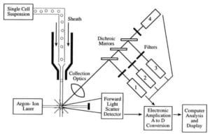
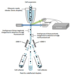
The properties measured include a particle’s relative size, relative granularity or internal complexity, and relative fluorescence intensity. Principle of flow cytometry the basic principle of flow cytometry is the passage of cells in single file in front of a laser so they can be detected, counted and sorted.

Immunologic Alterations In Ctla-4 Deficiency A And B Flow Cytometry Download Scientific Diagram
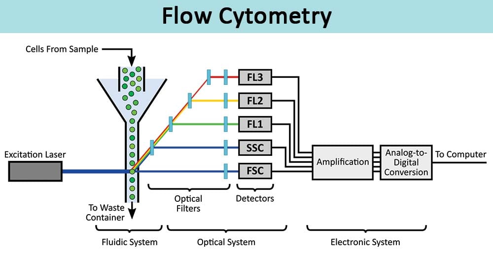
Flow cytometry » flow cytometry is the technical process that allows for the individual measurements of cell fluorescence and light scattering.

Flow cytometry test in hindi. Home test menu test directory: Flow cytometry this test is important in diagnosing cll. It uses a machine that looks for certain substances (markers) on or in cells that help identify what types of cells they are.
Limitations to flow cytometry include the facts that the laser can only analyze one cell at a time, cells. This test can be used to see if the lymphocytes in a sample of blood contain cll cells. Next working day if received before 1400 hrs.
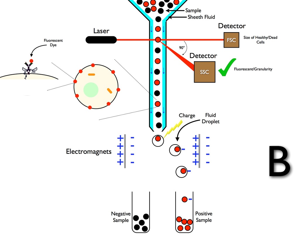
The diagnostic test for sepsis and inflammation based on the quantitation of cd64 expression levels on peripheral blood neutrophils is an example of a relative quantitative, flow cytometric method. Cell components are fluorescently labelled and then excited by the laser to emit light at varying wavelengths. » this information can be used to individually sort or separate subpopulations of cells.
Like tsne, spade extracts information across events in your data unsupervised and presents the result in a unique visual format. Prepare your cell suspensions for flow cytometry. Dna content analysis the measurement of cellular dna content by flow cytometry uses fluorescent dyes, such as propidium iodide, that intercalate into the dna helical structure.
Quantifying the pulse by measuring its height, area, and width. Flow cytometers utilize lasers as light sources to produce both scattered and fluorescent light signals that are read by detectors such as photodiodes or photomultiplier tubes. Flow cytometry basics guide | 7 principles of the flow cytometer fig.
Flow cytometry immunophenotyping is used primarily to help diagnose and classify blood cell cancers (leukemias and lymphomas) and to help guide their treatment. Flow cytometry is a laboratory method used to detect, identify, and count specific cells from blood, bone marrow, body fluids such as cerebrospinal fluid (csf), or tumors. This process is performed at rates of thousands of cells per second.
One of the most common applications is in the diagnosis of leukemia and lymphoma. Although most flow cytometry experiments involve labeling populations of cells that are relatively abundant, the number of cells required will vary depending upon the rarity of your cells. Whether you’re analyzing a sample looking for cancer cells, or separating stem cells in a laboratory, flow cytometry is the most effective method for counting and sorting heterogeneous fluids but its efficacy depends heavily on the machinery used.
Other flow cytometric methods in the clinical laboratory, such as leukemia lymphoma panels, are generally qualitative methods. Flow cytometry on fresh tissue and/or body fluid. F flow cytometry on fresh tissue and/or body fluid.
Flow cytometry often can detect recurrence of cancer before morphologic changes are detected. The rapidity of flow tests to identify abnormalities associated with specific diseases means that at least for the present and near future, flow cytometry will remain a viable technological. In general, researchers will stain between 1 x.
It may be used in follow up to a complete blood count (cbc) and wbc differential that show an increased number of lymphocytes or the presence of immature blood cells or other abnormal. However not all signals that are generated correspond to a particle of interest. For more information, contact the uf health pathology laboratories client services department at 888.375.labs (5227).
The fluorescent signal is directly proportional to the amount of dna in the nucleus and can identify gross gains or losses in dna. Flow cytometry is a technology that simultaneously measures and then analyzes multiple physical characteristics of single particles, usually cells, as they flow in a fluid stream through a beam of light. To avoid the processing of unwanted signals a.
Acute leukemia orientation panel the panel is designed to classify acute leukemia into all or aml. Overall, best practices for proper diagnosis should include clinical presentation, morphologic analysis, flow cytometry analysis, and other pertinent testing, such as cytogenetics ((wood et al., 2007). What do you know about this process and the reason why we use it?
Take the quiz now to let us know!
2

Neet Pg - Flowcytometry Introduction In Hindi Offered By Unacademy
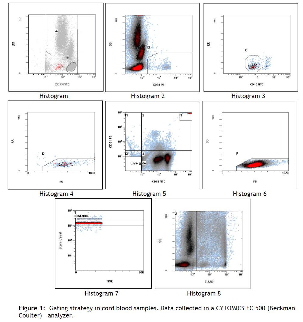
Direct Measurement Of Cd34 Cord Blood Stem Cell Absolute Counts Viability And Wbc Using Flow Cytometry Insight Medical Publishing

Storage Environment - Elabscience In 2020 Life Science Signal Transduction Cell Cycle

Marker Profile Analysis Of Mdscs A Flow Cytometry Revealed High Download Scientific Diagram
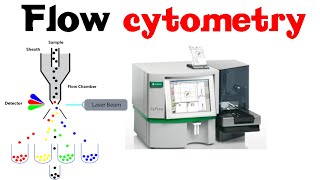
Flow Cytometry - Youtube
How Does Flow Cytometry Work Nanocellect
Flow Cytometry Principle How Facs Work - Shomus Biology
2

Direct Measurement Of Cd34 Cord Blood Stem Cell Absolute Counts Viability And Wbc Using Flow Cytometry Insight Medical Publishing
How Does Flow Cytometry Work Nanocellect

Introduction To Flow Cytometry Flow Cytometry Medical Technology Medical Laboratory Scientist
Flow Cytometry Principle How Facs Work - Shomus Biology

Flow Cytometry - Youtube
Flow Cytometry-definition Principle Parts Steps Types Uses

Guide To Gating In Flow Cytometry Bio-rad Flow Cytometry Medical Laboratory Science Science Infographics
Hematology White Blood Cells Lymph Nodes And Flow Cytometry Basicmedical Key

Flow Cytometry Principle Basics In Hindi - Part I - Youtube
Flow Cytometric Analysis Of Human Pancreatic Cancer Cell Line Download Scientific Diagram